
Personalized Eyewear Consultation by Appointment Only (215) 443-7706
- Home
- About
- Products
- Services
- Lens Lab
- Vision Care
- Newsletter
- Contact Us
Menu- Home
- About
- » Doug Wohl
- » Awards
- » Reviews
- Products
- » Bifocals
- » Computer Glasses
- » » Computer Vision Syndrome - Digital Eye Strain
- » Contact Lenses
- » » Are Contacts Right For You
- » » Guidelines for Contacts Wear and Care
- » » Instructions for Soft Contacts
- » Eyeglass Frames
- » » Frame Guide - Frame Shape and Face Shape
- » » Frame Materials
- » » Eyewear Maintenance
- » » Makeup Tips When Wearing Glasses
- » » Wiley X Eyewear Frames
- » Children Eyeglasses
- » » Eyewear for Babies and Young Children 0-5 Years Old
- » » Kids Corner
- » » 10 Tips for Buying Glasses for Children
- » Occupational Eyeglasses
- » Prescription Lenses
- » » Lens Coatings and Tints
- » » » Anti Reflective Coating
- » » Replacement Lens Options
- » Progressives
- » Readers
- » Safety Glasses
- » » Eye Injuries that Could be Prevented with Safety Glasses
- » » Prescription Safety Eye Glass Feature Guide
- » Single Vision Eyeglasses
- » Sports Eyewear
- » Sunglasses
- » » UV Eye Safety
- Services
- » Eye and Vision Exam
- » » Eye and Vision Problems
- » » Glaucoma Test
- » » Vision Refraction
- » Contact Lens Exam and Fitting
- » Eyeglass Repair
- Lens Lab
- Vision Care
- » Cornea
- » Detached Retina
- » Diabetic Retinopathy
- » Dry Eyes
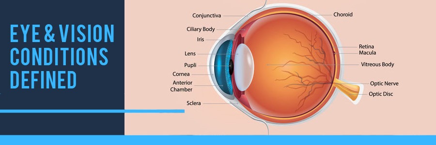
- » Eye and Vision Conditions
- » Fun Eye Facts
- » Eye Function and Parts Explained
- » FAQ
- » Macular Degeneration
- » Makeup Causing Eye Damage
- » Medication Side Effects to Vision
- » Optic Nerve Damage
- » Optical Terms and Definitions
- » Red Eye
- » Retina Damage
- Newsletter
- » Blue Light Blocker Glasses
- » Buying Glasses Online
- » Covid-19 Safety Practices
- Contact Us
- Home Vision Care Eye and Vision Conditions
Accommodation: Accommodation is the ability to adjust the focus of the eyes as the distance between the individual and the object changes. Children frequently use this vision skill in the classroom as they shift their attention (and focus) between their book and he chalkboard for sustained periods of time. Being able to maintain focus at near for sustained period of time is important for reading, writing and also taking tests.
Accommodative Dysfunction: Accommodative dysfunction simply means a focusing problem, particularly at near. This is not so much an eyesight (or clarity) difficulty as a problem in maintaining accurate, comfortable focus particularly with near work. Accommodative dysfunction is unrelated to aging changes in the lens of the eye. A child can fail to establish adequate focusing stamina during their early years of development, but in the clear majority of cases focusing dysfunction problems arise form fatigue as a result of sustained near visual tasks such as reading, writing, video games, computer use, etc. Prolonged near tasks can be fatiguing to some individuals. Problems typically manifest in the classroom as a student struggles to complete the assigned tasks within the time allotted for completion or while attempting to complete homework assignments. A student with accommodative dysfunction will become fatigued and have difficulty completing the assignments accurately and in a timely fashion.
Amblyopia (Lazy Eye): Amblyopia, commonly known as lazy eye, is an eye condition resulting in reduced vision and NOT correctable by glasses or contact lenses and not due to any eye disease. Amblyopia is a neurologically active process; the loss of vision takes place in the brain. If one eye sees clearly and the other is blurry, the brain can suppress (block or ignore) the eye with the blur. The brain can also suppress one eye to avoid double vision. The inhibition process (suppression) can result in a permanent decrease in the vision in the blurry eye that cannot be correct with glasses, lenses or lasik surgery.
Amblyopia is a visual defect that affects approximately 2 or 3 out of every 100 children in the United States. Three percent (3%) of children under six years of age have some form of amblyopia. Amblyopia is the loss or lack of development of clear vision in just one eye which cannot be corrected by glasses or contact lenses. The result is often a loss of stereoscopic vision (3D) and depth perception.
Early detection and treatment offer the best outcome. Scientific research by the National Eye Institute, National Institutes of Health, and Department of Health and Human Services has now proven that effective treatment can take place up to the age of 17. Scientific research on treatment after the age of 17 has not yet been done.
Treatment of amblyopia involves therapeutic glasses, drops, vision therapy, or patching. The length of the treatment period for Amblyopia does increase dramatically the longer the condition has existed prior to treatment, so early detection and treatment are vital.
Astigmatism: Astigmatism is a vision condition that causes blurred vision due either to the irregular shape of the cornea, the clear front cover of the eye, or sometimes the curvature of the lens inside the eye. An irregular shaped cornea or lens prevents light from focusing properly on the retina, the light sensitive surface at the back of the eye. As a result, vision becomes blurred at any distance.
The curvature of the cornea and lens causes light entering the eye to be bent in order to focus it precisely on the retina at the back of the eye. In astigmatism, the surface of the cornea or lens has a somewhat different curvature in one direction than another. In the case of the cornea, instead of having a round shape like a basketball, the surface of the cornea is more like a football in shape. Thus, the eye is unable to focus light rays to a single point causing vision to be out of focus at any distance.
Astigmatism is a very common vision condition. Most people have some degree of astigmatism. Slight amounts of astigmatism usually don’t’ affect vision. However, larger amounts of astigmatism cause distorted or blurred vision, eye discomfort and headaches.
Behavioral Optometrist: see Developmental Optometrist below
Binocular Fusion: Binocular fusion refers to the brain’s ability to gather information received from each eye separately and form a single, unified image. A child’s eyes must be precisely aligned or blurred or double vision, discomfort, confusion or avoidance may result.
If that occurs, the brain often subconsciously suppresses or inhibits the vision in one eye to avoid confusion. That eye may then develop poorer visual acuity (amblyopia or lazy eye).
Binocular Vision: Binocular vision is the result of both eyes working together as a team; both eyes work together smoothly, accurately, equally and simultaneously. Binocular vision literally means both eyes at once.
The process of binocular vision reflects complex interactions within the eye-brain continuum. For neural binocular summation to occur, inputs from both eyes to the brain must be synchronized in both space and time. Alignment of the eyes is maintained via ongoing collaboration of eyes and brain. Binocular dysfunctions such as suppression and anomalous correspondence demonstrate cortical adaptations in the eye-brain function to minimize visual confusion and maintain some level of visual performance.
Binocular Depth Perception: Binocular depth perception is a result of binocular vision and the result of successful eye teaming and stereoscopic vision; the ability to visually perceive three dimensional space; the ability to visually judge relative distances between objects; a visual-motor skill that aids accurate movement in three-dimensional space.
Binocular Vision Impairment: Binocular vision impairment is a visual defect in which the two eyes fail to work together as a coordinated team resulting in a partial or total loss of binocular depth perception and stereoscopic vision. Binocular vision impairment affects at least 12% of the population. Amblyopia and strabismus are the most commonly known types of binocular vision impairment.
Blepharitis: Blepharitis is an inflammation of the eyelids causing red, irritated, itchy eyelids and the formation of dandruff-like scales on eyelashes. It is a common eye disorder caused by either bacteria or a skin condition such as dandruff of the scalp or acne rosacea. It affects people of all ages. Although uncomfortable, blepharitis is not contagious and generally does not cause nay permanent damage to eyesight.
Anterior blepharitis occurs at the outside front edge of the eyelid where the eyelashes are attached. Anterior blepharitis is commonly caused by bacteria (staphylococcal blepharitis) or dandruff of the scalp and eyebrows (seborrheic blepharitis). It may also occur due to a combination of factors, or less commonly may be the result of allergies or an infestation of the eyelashes.
Posterior blepharitis affects the inner edge of the eyelid that comes in contact with the eyeball. Posterior blepharitis can be caused by irregular oil production by the glands of the eyelids (meibomian blepharitis) which create a favorable environment for bacterial growth. It can also develop as a result of other skin conditions such as acne rosacea and scalp dandruff.
Cataracts: Cataracts are cloudy or opaque areas in the normally clear lens of the eye. Depending upon their size and location, cataracts can interfere with normal vision. Usually cataracts develop in both eyes, but one may be worse than the other. Cataracts can cause a decrease in contrast sensitivity, a dulling of colors and increased sensitivity to glare.
Chalazion: A chalazion, also known as a meibomian gland lipogranuloma, is a cyst in the eyelid that is caused by inflammation of a blocked meibomian gland, usually on the upper eyelid. A chalazion is a slowly developing lump that forms due to blockage and swelling of an oil gland in the eyelid. Chalazions differ from styles (hordeolums) in that they are usually painless apart from the tenderness caused when they swell.
Color Vision Deficiency (Color Blindness): Color vision deficiency is the inability to distinguish certain shades of colors or, in more severe cases, see colors at all. It is most often of genetic nature, but may also occur because of eye, nerve, brain damage, or due to exposure to certain chemicals. Other causes are Shaken Baby Syndrome (this can cause retina and brain damage and therefore cause color blindness), accidents and other trauma (swelling of the brain in the occipital lobe), and UV damage to the retina.
Various types of color vision deficiencies or color blindness:
Dichromacy is a moderately severe color vision defect in which one of the three basic color mechanisms is absent or not functioning. It is hereditary and sex-linked, predominantly affecting males. Dichromacy occurs when one of the cone pigments is mission and color is reduced to two dimensions.
Deuteranopia consists of impairment in perceiving medium wavelengths, such as greens. Deuteranomaly is a less severe form of deuteranopia. Those living with deuteranomaly cannot see reds and greens; however they can still distinguish them in most cases.
Protanopia is a severe form of red-green color-blindness, where there is impairment in perception of very long wavelengths, such as reds. To these sufferers, reds are perceived as beige and greens tend to look like reds. Protanomaly is a less severe version.
Computer Vision Syndrome (CVS): Computer vision syndrome describes a group of eye and vision-related problems that result from prolonged computer use. Many individuals experience eye discomfort and vision problems when viewing a computer screen for extended period.
The extent to which individual’s experience computer vision syndrome often depends on the level of their visual abilities and the amount of time spent looking at the computer screen. Uncorrected vision problems like farsightedness and astigmatism, inadequate eye focusing or eye coordination abilities, and again changes of the eyes, such as presbyopia, can all contribute to the development of computer vision syndrome.
Eyeglasses or contact lenses prescribed for general use are often not adequate for computer use. Specific occupational lenses prescribed to meet the unique demands of computer work may be needed. Special lens designs, lens powers or lens tints or coatings may help to maximize visual abilities and comfort.
Conjunctivitis: Conjunctivitis is an inflammation or infection of the conjunctiva, the thin transparent layer of tissue that lines the inner surface of the eyelid and covers the white part of the eye. Conjunctivitis, often called “pink eye”, is a common eye disease, especially in children. It may affect one or both eyes. Some forms of conjunctivitis can be highly contagious and easily spread in schools and at home. While conjunctivitis is usually a minor eye infection, sometimes it can develop into a more serious problem which is why it is recommended that individuals with conjunctivitis seek medical treatment.
Conjunctivitis may be caused by a viral or bacterial infection. It can also occur due to an allergic reaction to irritants in the air like pollen and smoke, chlorine in swimming pools, and ingredients in cosmetics or other products that come in contact with the eyes. Sexually transmitted diseases like Chlamydia and gonorrhea are less common causes of conjunctivitis.
Various types of conjunctivitis:
Allergic Conjunctivitis: occurs more commonly among people who already have seasonal allergies. At some point they come into contact with a substance that triggers an allergic reaction in their eyes.
Bacterial Conjunctivitis: is an infection most often caused by staphylococcal or streptococcal bacteria from your own skin or respiratory system. Infection can also occur by transmittal from insects, physical contact with other people, poor hygiene (touching the eye with unclean hands), or by use of contaminated eye makeup and facial lotions.
Chemical Conjunctivitis: treatment for chemical conjunctivitis requires careful flushing of the eyes with saline and may require topical steroids. The more acute chemical injuries are medical emergencies, particularly alkali burns, which can lead to severe scarring, intraocular damage or even loss of an eye.
Giant Papillary Conjunctivitis: is a type of allergic conjunctivitis caused by the chronic presence of a foreign body in the eye. This condition occurs predominantly with people who wear hard or rigid contact lenses, wear soft contact lenses that are not replaced frequently, have an exposed suture on the surface of the eye, or have a glass eye.
Ophthalmia Neonatorum: is a severe form of bacterial conjunctivitis that occurs in newborn babies. This is a serious condition that could lead to permanent eye damage unless it is treated immediately. Ophthalmia neonatorum occurs when an infant is exposed to Chlamydia or gonorrhea while passing through the birth canal.
Viral Conjunctivitis: is most commonly caused by contagious viruses associated with the common cold. The primary means of contracting this is through exposure to coughing or sneezing by persons with upper respiratory tract infections. It can also occur as the virus spreads along the body’s own mucous membranes connecting lungs, throat, nose, tear ducts, and conjunctiva.
Corneal Ulcer: see Keratitis below
Convergence: Convergence is the ability to turn the two eyes toward each other to look at a close object. School deskwork is one instance in which a child depends on this vision skill.
Convergence Insufficiency: An eye coordination problem in which the eyes have a tendency to drift outward when reading or doing close work. This is a condition in which the muscles of the eye responsible for convergence (turning the eyes in) appear to be weak, at least relative to the muscles responsible for divergence (turning the eyes out).
Convergence insufficiency is a common condition that is characterized by a person’s inability to maintain proper binocular eye alignment on objects as they approach from distance to near. This results in “strained” eyes and the individual complains of headache, eyestrain, blurred vision, or fatigue with continued efforts at near work.
Corneal Abrasion: A cut or scratch on the cornea, the clear front cover of the eye. Common causes of corneal abrasions include fingernails, mascara wands, paper cuts, tree branches, animal scratches, cigarettes, inverted eyelashes, and blunt trauma to the eye.
Pain, irritation, tearing, red eye, twitching of the eye, decreased vision, and sensitivity to light are common complaints that accompany a corneal abrasion. If there is inflammation inside the eye, a dull ache may be felt inside the eye.
If a corneal abrasion is not treated appropriately, scarring and ulceration of the cornea are possible. Treatment of a corneal abrasion with over the counter (OTC) drugs advertised to decrease redness should not be used to self treat corneal abrasions. These drugs act by constricting the blood vessels in the eye, decreasing the blood supply to the eye and delaying healing. Also, the preservatives in these drops may further irritate the cornea.
Crossed Eyes: see Strabismus below
Cumulative Deficit: Cumulative deficit is the tendency for a deficit between achievement and grade placement to accumulate over the years of school such that secondary students with disabilities demonstrate much larger deficits than students in the lower grades with the same disabilities.
Developmental Optometrist (also known as a Behavioral Optometrist): Developmental Optometrists are interested in how the eyes work together with the brain and the rest of the body to perceive images and help us navigate the world. It is an extension of the work of more traditional ophthalmologists and optometrists who specialize in how the eyes receive images, and who typically prescribe glasses, contact lenses, medicine, or surgery. A Developmental Optometrist is a doctor of optometry who belongs to an international branch of optometry which specializes in the practice of developmental vision and vision therapy. Developmental Optometrists consider how environmental, nutritional and/or behavioral factors affect visual health; therefore, the term “behavioral” is used. Developmental Optometrists also evaluate the process of development and the proper use of age appropriate vision skills as well as sensory integration processes.
Diabetic Retinopathy (Diabetes): Diabetic retinopathy is a condition occurring in persons with diabetes, which causes progressive damage to the retina, the light sensitive lining at the back of the eye.
People with Type 1 or Type 2 diabetes are at risk for the development of diabetic retinopathy. Hispanic and African Americans are at greater risk for developing diabetic retinopathy. People with other medical conditions such as high blood pressure and high cholesterol are at a greater risk. Pregnant women face a higher risk of developing diabetes and diabetic retinopathy. If gestational diabetes develops, the individual is at much higher risk of developing diabetes as they age. Diabetic retinopathy is a serious sight-threatening complication of diabetes.
Per the American Diabetes Association, an estimated 54 million Americans aged 40 to 74 (40.1% of the US population in this age group) have pre-diabetes, a condition that puts them at high risk for developing type 2 diabetes. 21 million Americans have diabetes and another 6 million have diabetes but are unaware that they have the disease.
A dilated eye exam is the first line of detection for diabetes, since the eye is the only place on the body that blood vessels can be seen without having to look through the skin.
Diabetic retinopathy is the result of damage to the tiny blood vessels that nourish the retina. They leak blood and other fluids that cause swelling of retinal tissue and clouding of vision. The condition usually affects both eyes. The longer a person has diabetes, the more likely they will develop diabetic retinopathy. If left untreated, diabetic retinopathy can cause blindness.
Various types of diabetic retinopathy:
Non-proliferative diabetic retinopathy (NPDR): is the early sate of the disease in which symptoms will be mild or non-existent. In NPDR, the blood vessels in the retina are weakened causing the tiny bulges called microaneurysms to protrude from their walls. The microanuerysms may leak fluid into the retina, which may lead to swelling of the macula.
Proliferative diabetic retinopathy (PDR): is the more advanced form of the disease. At this stage, circulation problems cause the retina to become oxygen deprived. As a result new fragile blood vessels can begin to grow in the retina and into the vitreous, the gel-like fluid that fills the back of the eye. The new blood vessel may leak blood into the vitreous, clouding vision. Other complications of PDR include detachment of the retina due to scar tissue formation and the development of glaucoma. Glaucoma is an eye disease defined as progressive damage to the optic nerve. In cases of proliferative diabetic retinopathy, the cause of this nerve damage is due to extremely high pressure in the eye. If left untreated, proliferative diabetic retinopathy can cause severe vision loss and even blindness.
Diplopia (also known as Double Vision): Double vision (diplopia) that is only evident when looking through both eyes and disappears if one eye is closed or covered. The condition is caused by misalignment of the eyes by the extraocular muscles (the muscles around the eyeball that control gaze). This may be due to strabismus (misalignment of the eyes from birth), neurologic damage to the extraocular muscles (as from a brain abscess, stroke, and head trauma or brain tumor), diabetes, myasthenia gravis, Graves’s disease, or trauma to the yes muscles, as from a fracture of the orbit.
Treatment of double vision consists of vision therapy, surgical straightening of the eye or a combination of the two. Vision therapy is aimed at re-aligning the misaligned eye where possible without surgery and re-stimulating the part of the visual pathway to the brain which is not working correctly.
Drusen: Drusen are small yellow or off-white deposits that form either in the tissue layer underneath the retina or on the top of the optic nerve head. Though the exact cause of macular drusen is still unknown, their appearance near the macula is one of the most common signs of macular degeneration development. Drusen that develop away from the macula are typically considered safe and are not indicative of future vision impairment.
While the presence of drusen near the macula doesn’t necessarily indicate macular degeneration, it does mean that the eye may be at risk. Drusen can even be present in the eye for years without impairing vision at all. The presence of macular drusen can be detected during a regular, dilated eye examination.
Dry Eye: Dry eye is a condition in which there are insufficient tears to lubricate and nourish the eye. Tears are necessary for maintaining the health of the front surface of the eye and for providing clear vision. People with dry eyes either do not produce enough tears or have a poor quality of tears. Dry eye is a common and often chronic problem, particularly in women, older adults or due to some prescription medications.
Tears are made up of three layers: oil, water, and mucus. Each component serves a function in protecting and nourishing the front surface of the eye. A smooth oil layer helps to prevent evaporation of the water layer, while the mucin layer functions in spreading the tears evenly over the surface of the eye. If the tears evaporate too quickly or do not spread evenly over the cornea due to deficiencies with any of the three tear layers, dry eye symptoms develop.
Eye drops to treat dry eye are best prescribed by an eye doctor. There are a variety of prescription and OTC (over the counter) eye drops available. Each type of eye drop treats either the oil, water or mucus layer. It is important to use the specific type of eye drop developed to treat the particular type of dry eye.
Dyslexia (Dyslexic): Developmental dyslexia is a selective impairment of reading skills despite normal intelligence, sensory acuity, and instruction. Several perceptual studies have suggested that dyslexic individuals process visual information more slowly than normal individuals. Visual abnormalities are reported to be found in more than 75% of reading-disabled individuals. Therefore, it is important to rule out problems with sensory integration and/or sensory processing (including visual acuity and visual processing) before labeling an individual as dyslexic.
Various types of dyslexia:
Dysnemkinetic (visual and memory related dyslexia): Deficit in the ability to develop the necessary motor skills for writing symbols such as letters or numbers as the neural skills are not imprinted adequately in the memory of the brain.
Dysphonetic (auditory dyslexia): Deficit in the ability to correctly associate the sound of a given letter or letter combination and to differentiate between nuances of sounds.
Dyseidetic: Deficit in the ability to recognize words as a whole and to pronounce them correctly.
Dysphoneidetic: Deficit in the ability to correctly associate the sound of a given letter or letter combination within a word as a whole, and to pronounce them correctly.
Dysnemkinphonetic: Deficit in the ability to develop the necessary motor skills for writing letters or numbers and in pronouncing the symbols correctly.
Dysnemkineidetic: Deficit in the ability to develop the necessary motor skills for writing letters or numbers and in pronouncing the symbols correctly either by syllable or by the word in its entirety.
Dysnemkinphoneidetic: Deficit in the ability to develop the necessary motor skills for writing letters or numbers and in pronouncing the symbols correctly either by syllable or by the word in its entirety.
Farsightedness: see Hyperopia below
Field of Vision: Field of vision is the wide area over which vision is possible. It is important that an individual be aware of objects in the periphery (left and right sides and up and down) as well as in the center of the field of vision. Near central or para-central vision is important for reading ability.
Figure-Ground: One part of a perceptual configuration stands out while the remainder forms a background. Some people have difficulty in separating the configurations which may cause academic problems.
Floaters (Spots): Floaters are small, semi-transparent or cloudy specks or particles within the vitreous, which is the clear, jelly-like fluid that fills the inside of your eyes. They appear as specks of various shapes and sizes, threadlike strands or cobwebs. Because they are within your eyes, they move as your eyes move and seem to dart away when you try to look at them directly.
Sports are often caused by small flecks of protein or other matter trapped during the formation of your eyes before birth. They can also result from deterioration of the vitreous fluid, due to aging, or from certain eye diseases or injuries.
Floaters or spots can be indications of a serious problem, and you should have a dilated eye exam to evaluate the health of your eyes.
Fluctuating Vision: If you experience frequent changes in how clearly you can see, it may be a sign of diabetes or hypertension (high blood pressure). These chronic conditions can damage the tiny blood vessels in the retina, the light sensitive layer at the back of the eye, causing vision loss that can sometimes be permanent.
Glaucoma: Glaucoma is often called “the sneak thief of sight” because it can strike without pain or other symptoms and is one of the leading causes of blindness in the United States. Unfortunately, the vast majority of Americans – 91% - incorrectly believe glaucoma is preventable. Although glaucoma is not preventable, if diagnosed and treated early, doctors of optometry can help a patient control the disease. Medication or surgery can slow or prevent further vision loss. However, vision already lost to glaucoma cannot be restored.
Glaucoma is diagnosed through a comprehensive eye examination. To establish a diagnosis of glaucoma, several factors must be present: Because glaucoma is a progressive disease, meaning it worsens over time, a change in the appearance of the optic nerve, a loss of nerve tissue, and a corresponding loss of vision confirm the diagnosis. Some optic nerves have a suspicious appearance, resembling nerves with glaucoma, but the patients may have no other risk factors or signs of glaucoma. These patients should be closely followed with routine, annual comprehensive eye exams to monitor for change.
Glaucoma is a group of eye disorders leading to progressive damage to the optic nerve, and is characterized by loss of nerve tissue resulting in loss of vision.
Various types of glaucoma:
Primary open-angle glaucoma: This is the most common form of glaucoma. One theory is that glaucoma is thought to develop when the eye’s drainage system becomes inefficient over time. This leads to an increased amount of fluid and a gradual buildup of pressure within the eye. Other theories of the cause of the optic nerve damage include poor perfusion, or blood flow, to the optic nerve. Damage to the optic nerve is slow and painless and a large portion of vision can be lost before vision problems are noticed.
Angle-closure glaucoma: This type of glaucoma, also called closed-angle glaucoma or narrow angle glaucoma, is a less common form of the disease. It is a medical emergency that can cause vision loss within a day of its onset. Angle-closure glaucoma can be chronic (progressing gradually) or acute (appearing suddenly). The acute form occurs when the iris completely blocks the drainage of the aqueous fluid. Although an acute attack often affects only one eye, the other eye may be at risk of an attack as well.
Acute narrow-angle glaucoma occurs suddenly, when the colored portion of your eye (iris) is pushed or pulled forward. This causes blockage of the drainage angle of the eye, where the trabecular meshwork allows outflow of fluids.
When internal eye structures are blocked in this way, your eye's internal pressure (intraocular pressure or IOP) may spike and possibly damage the optic nerve that transmits images from the eye to the brain.
Acute angle-closure (closed-angle or narrow-angle) glaucoma produces symptoms such as eye pain, headaches, halos around lights, dilated pupils, vision loss, red eyes, nausea and vomiting.
These signs may last for hours or until the IOP is reduced. With each narrow-angle glaucoma attack, part of your peripheral vision may be lost.
Acute angle-closure glaucoma is a medical emergency. If the high eye pressure is not reduced within hours, it can cause permanent vision loss. Anyone who experiences these symptoms should contact an ophthalmologist immediately or go to a hospital emergency room.
Secondary glaucoma: This type of glaucoma occurs as a result of an injury or other eye disease. It may be caused by a variety of medical conditions, medications, physical injuries, and eye abnormalities. Infrequently, eye surgery can be associated with secondary glaucoma.
Normal-tension glaucoma: In this form of glaucoma, eye pressure remains within what is considered to be the “normal” range, but the optic nerve is damaged nevertheless. Why this happens is unknown.
Hyperopia (Farsightedness): Farsightedness, or hyperopia, as it is medically termed, is a vision condition in which distant objects are usually seen clearly, but close ones do not come into proper focus. Farsightedness occurs if your eyeball is too short or the cornea has too little curvature, so light entering your eye is not focused correctly.
Common signs of farsightedness include difficulty in concentrating and maintaining a clear focus on near objects, eye strain, fatigue and/or headaches after close work, aching or burning eyes, irritability or nervousness after sustained concentration.
Common vision screenings, often done in schools or at the pediatricians’ office, are generally ineffective in detecting farsightedness.
Keratitis: Corneal ulcers (keratitis) occur after corneal trauma with a foreign body (including contact lenses), and with dry eyes or lid disease which allow bacteria or fungi to enter the cornea, causing a deep infection and inflammation. This condition may cause severe pain, reduce visual clarity, produce a corneal discharge, and perhaps erode the cornea. As a rule, the deeper the corneal infection, the more severe the symptoms and complications. Microbial infections such as keratitis are the most serious complication of contact lens wear.
Keratoconus: Keratoconus is a vision disorder that occurs when the normally round cornea (the front part of the eye) becomes thin and irregular (cone) shaped. This abnormal shape prevents the light entering the eye from being focused correctly on the retina and causes distortion of vision.
Symptoms usually appear in the late teens or late 20’s. Keratoconus may progress for 10 to 20 years and then slow in its progression. Each eye may be affected differently.
As keratoconus progresses, the cornea bulges more and vision becomes more distorted. In a small number of cases, the cornea will swell and cause a sudden and significant decrease in vision. The swelling may last for weeks or months as the crack heals and is gradually replaced by scar tissue.
Eyeglasses or soft contact lenses may be used to correct the mild nearsightedness and astigmatism that is caused by the early stages of keratoconus. As the disorder progresses and the cornea continues to thin and change shape, rigid gas permeable contact lenses can be prescribed to correct vision adequately. The contact lenses must be carefully fitted and frequent checkups and lens changes may be needed to achieve and maintain good vision.
In a few cases, a corneal transplant is necessary. However, even after a corneal transplant, eyeglasses or contact lenses are often still needed to correct vision.
Lazy Eye: see Amblyopia above
Learning Related Vision Problems: Vision disorders that interfere with reading and learning. A learning-related visual problem directly affects how we learn, read, or sustain close work. Visual problems in any of the following areas can have a significant impact on learning:
● eye tracking skills – eyes following a line of print
● eye teaming skills – two eyes working together as a synchronized team
● binocular vision – simultaneously blending the images from both eyes into one Image
● accommodation – eye focusing
● visual-motor integration – eye-hand coordination
● visual perception – visual memory, visual form perception, and visualization
As vision and learning are intimately connected, a vision problem can be easily mistaken for a learning problem. Children with visual problems can be misdiagnosed as having Learning Disabilities, ADHD, or Dyslexia.
When parents observe that their child is struggling in school, it’s time to have their vision properly evaluated with a comprehensive learning related eye examination.
Macular Degeneration: Macular degeneration is defined as a disease that gradually destroys the central area of the retina, known as the macula. The macula transforms light waves from directly in front of the eye into nerve signals that the brain processes into discernable images. When the macula becomes damaged, crisp central vision is affected.
Because macular degeneration affects only straight-ahead vision, it cannot lead to total blindness. It can, however, severely impair the ability to perform day to day activities such as reading or driving. Macular degeneration is painless, and in the early stages, the brain easily compensates for vision loss, particularly if macular degeneration is restricted to one eye.
Macular degeneration is the leading cause of severe vision loss and blindness in people over the age of 65. It is commonly referred to as age-related macular degeneration, or ARMD. A variety of factors influence the risk of developing macular degeneration: Age, gender (females are at greater risk than males), lighter colored eyes, genetic history, smoking, diets high in saturated fat and cholesterol, low dietary consumption of antioxidants, high-blood pressure, higher than normal levels of cholesterol and obesity.
One in six Americans develops AMD between the ages of 55 and 64 and one in three Americans over 75 has AMD. About 10% of those with AMD eventually suffer severe vision loss. The incidence of AMD is expected to triple by 2025, as the population ages.
Various types of macular degeneration:
Dry Macular Degeneration: dry macular degeneration accounts for nearly 90% of ARMD cases. This type of macular degeneration slowly deteriorates central vision over a long period of time. Drusen that appear during the gradual development of dry ARMD may have no effect on an individual’s vision, making awareness of dry ARMD’s earliest phases difficult. Vision impairment due to dry ARMD can often be stable for years before progressing any further.
Wet Age-Related Macular Degeneration: wet macular degeneration is the less common variety of ARMD, tends to develop quickly and suddenly, causing rapid vision loss. Wet ARMD causes new blood vessels to form under the retina. Since these new vessels are delicate, they soon begin to leak blood and fluid, distorting the macula and compromising sharp central vision.
Juvenile Macular Degeneration (JMD): JMD is used to describe a group of inherited disorders affecting children and younger adults. Examples of these include: Best’s disease, Doyne’s honeycomb retinal dystrophy, Sorsby’s disease, Stargardt’s disease.
Cystoid Macular Degeneration: cystoid macular degeneration is the development of fluid-filled cysts (sacs) in the macular region, associated with aging, inflammation, or severe myopia (nearsightedness).
Diabetic Macular Degeneration: see Diabetic Retinopathy
Retinal Pigment Epithelial Detachment: This is a rare form of wet macular degeneration in which fluid leakage from the choroid causes the detachment or disappearance of the pigmented retinal epithelium.
Though dry age related macular degeneration is the less severe type of ARMD, it currently has no known treatments or cure. There are treatments for wet ARMD, including macular degeneration surgery and medication.
Monovision: Monovision is a treatment technique that is often prescribed for people age 40 and older who are affected by presbyopia. Presbyopia occurs when, as part of the natural aging process, the eye’s crystalline lens loses its ability to bring close objects into clear focus.
Monovision means wearing a contact lens for near vision on one eye and, if needed, a lens for distance vision on the other eye. Monovision can also be accomplished with refractive surgery, laser vision correction commonly known as LASIK.
Alternative treatments for presbyopia include a combination of contact lenses and reading glasses, multifocal spectacle lenses or multifocal contact lenses.
Myopia (Nearsightedness): Myopia, as it is medically termed, is a vision condition in which close objects are seen clearly, but objects farther away appear blurred. Nearsightedness occurs if the eyeball is too long or the cornea, the clear front cover of the eye, has too much curvature. Thus, the light entering the eye isn’t focused correctly on the retina and distant objects look blurred.
Myopia is a very common vision condition affecting nearly 30% of the U.S. population. Some research supports the theory that nearsightedness is hereditary. There is also growing evidence that it is influenced by the visual stress of too much close work.
Generally, myopia first occurs in school-age children. Because the eye continues to grow during childhood, it typically progresses until about age 20. However, myopia may also develop in adults due to visual stress or health conditions such as diabetes.
Some people may experience blurred distance vision only at night. This “night myopia” may be due to the low level of light making it difficult for the yes to focus properly or the increased pupil size during dark conditions, allowing more peripheral, unfocused light rays to enter the eye.
People who do an excessive amount of near vision work may experience false or “pseudo” myopia. Their blurred distance vision is caused by over use of the eyes’ focusing mechanism. After long periods of near work, their eyes are unable to refocus to see clearly in the distance. The symptoms are usually temporary and clear distance vision may return after resting the eyes. However, over time constant visual stress may lead to a permanent reduction in distance vision.
Nearsightedness: see Myopia above
Nystagmus: Nystagmus is a vision condition in which the eyes make repetitive, uncontrolled movements, often resulting in reduced vision. Vertical nystagmus occurs much less frequently than horizontal nystagmus and is often, but not necessarily, a sign of serious brain damage. Nystagmus can be a normal physiological response or a result of a medical problem.
There are many causes of nystagmus. Nystagmus may be present at birth. It may be a result of the lack of development of normal binocular fixation early in life. Some other conditions that nystagmus may be associated with include: albinism, optic atrophy, color blindness, very high nearsightedness, severe astigmatism, acute labyrinthitis (inflammation of the inner ear), brain lesions, alcohol, medications, and multiple sclerosis.
Nystagmus is a symptom, not a disease. If abnormal, it indicates a medical problem.
Ocular Allergies: Ocular allergies, also known as allergic conjunctivitis, is the inflammation of the conjunctiva (the membrane covering the white part of the eye) due to allergy. An allergy is the abnormal response of sensitive eyes to contact with allergens and other irritating substances. Symptoms consist of redness, edema of the conjunctiva, itching and increased tears or watery eyes.
Treatment of allergic conjunctivitis is to avoid the allergen (dust, pollen, grasses, pets, etc) and treatment with antihistamines, either in the form of eye drops or systemic in the form of tablets.
Be cautious if choosing to use over the counter (OTC) medications. Many OTC medications contain chemicals and preservatives which can further irritate the eyes. There are homeopathic eye drops as well as prescription eye drops that are recommended.
Ocular Hypertension: Ocular hypertension refers to any situation in which the pressure inside the eye, called intraocular pressure, is higher than normal. Ocular hypertension should not be considered a disease by itself. Instead ocular hypertension is a term that is used to describe individuals who should be observed more closely than the general population for the onset of glaucoma.
The necessity of observation is to evaluate the nerve fiber layer within the eye. Up to 40% of the nerve fiber tissue can be lost before any visual field loss is noticed by the patient. The preferred methods to evaluate the nerve fiber layer is with dilated eye exams and testing using an OCT.
Ocular Migraine: An ocular migraine, typically, is not cause for concern. However, ocular migraine symptoms can interfere with daily activities such as reading, using the computer or driving. These episodes, also called migraine aura or optical migraine, occur without a headache and are relatively common. Symptoms of an ocular migraine include: Flashes of light, zigzagging patterns, blind spots, shimmering spots or stars.
The causes of an ocular migraine are unknown. Some individuals relate ocular migraines to eating certain foods such as chocolate, nuts, shellfish or artificial sweeteners. Other people experience ocular migraines after drinking caffeine or alcohol. Other people believe it is tension or fatigue. Researchers have not found a consistent link to the cause of ocular migraines.
If ocular migraines persist, worsen or become accompanied by a migraine headache it is time to schedule an appointment with the eye doctor.
Ophthalmologist or Pediatric Ophthalmologist: An ophthalmologist is a doctor of medicine (M.D.) specializing in surgery and diseases of the eye. The term ophthalmologist implies a medically trained surgical specialist. Since ophthalmologist perform operations on eyes, they are generally categorized as surgeons and as a rule they are under informed about subject areas such as: Visual processing, convergence, accommodation and vision therapy. Some ophthalmologists concede this. In the medical journal, Transaction of the American Ophthalmological Society, eye muscle surgeon and researcher David Guyton, M.D., states: “We (ophthalmologists) have probably abdicated the study of accommodation and convergence to the optometric profession. A perusal of the literature will reveal that most of the advances in this area are being made in the optometric institutions by vision scientists who use definitions and terms with which we are not familiar.”
Optometrist: An optometrist is an eye doctor (O.D.) trained as a primary care optometric physician. Optometrists diagnose and treat eye diseases such as glaucoma and retinal diseases; diagnose systemic (body wide) conditions such as hypertension and diabetes that may affect the eyes; examine, diagnose and treat visual conditions such as nearsightedness, farsightedness, astigmatism and presbyopia; prescribe glasses, contact lenses, vision rehabilitation and medications as well as perform minor surgical procedures such as the removal of foreign bodies.
Orthoptic Therapy: Orthoptic therapy is a limited form of vision therapy which only trains eye teaming skills and visual acuity and does not treat other visual dysfunctions. Orthoptic therapy does not address visual perception, visual processing, visual memory or sensory integration.
Perception: Visual perception is the total process responsible for the reception and understanding of what is seen. Good visual perception is necessary for successful school achievement.
Form perception is the ability to organize and recognize visual images as specific shapes. The shapes the child encounters are remembered, defined and recalled when development of reading skills begin.
Perceptual Abilities: Perceptual abilities are the abilities to process, organize, and interpret the information obtained by the five senses; a function of the brain.
Perceptual Speed: The specific meaning of the term perceptual speed varies, depending upon the way a given test measures this ability. May refer to motor speed, how fast something is copied or manipulated, or to visual discrimination, e.g., how quickly identical items in a given series are identified.
Pinguecula: A pinguecula is a raised growth on the eye that appears as a small, yellowish lesion. It can appear on either side of the cornea, but usually appears on the nasal side. Pingueculas are more common in people who spend time in direct sunlight. Welding is a significant occupational risk.
In some cases, pingueculae become swollen and inflamed, a condition called pingueculitis. Irritation and eye redness from pingueculitis usually result from exposure to sun, wind, dust or extremely dry conditions. A pinguecula does not usually require treatment, but surgery may be necessary for cosmetic reasons. Protecting eyes from UV radiation is a preventive measure for this condition.
Presbyopia: Presbyopia is a progressive condition that is a natural part of aging. Presbyopia is a decrease in the ability to focus sharply on nearby objects and often results in the need to use magnifying reading glasses, bifocals or progressive lenses.
Presbyopia typically begins between the ages of 40 and 45. Initially reading materials or small objects must be moved farther away from our eyes and brighter light must be used for reading. Presbyopia affects either myopic (nearsighted) or hyperopic (farsighted) eyes, but shows its symptoms earlier in people with hyperopia. People that have never had to wear spectacles will be affected. The most significant symptom is a rapid deterioration of near vision.
Pterygium: A pterygium is a raised, wedge or wing shaped growth of benign fibrous tissue with blood vessels, typically located on the sclera. Pterygia may grow onto the eye’s cornea and interfere with vision. A pterygium resembles tissue or film growing over the eye. Pterygia are most often directly related to over-exposure to the sun. Dry, dusty conditions may also be a factor.
Treatment depends on the pterygium’s size and the symptoms it causes. Lubricants or mild steroid eye drops may be prescribed to reduce swelling and redness. In some cases, surgical removal of the pterygium is necessary.
Ptosis: Ptosis is an abnormal drooping of the upper eyelid. The drooping may worsen after being awake longer because the eyelid muscles tire. If severe enough and left untreated, the drooping eyelid can cause other conditions like amblyopia or astigmatism.
Ptosis occurs when the muscles that raise the eyelid are not strong enough to hold the eyelid up in a normal position. Ptosis can affect one eye or both eyes and is more common in the elderly, as muscles in the eyelids begin to deteriorate.
Reading Comprehension: To gain comprehension throughout the reading process, we are constantly taking in the visual information and decoding it from the written word into a mental image. Memory and visualization are also used to constantly relate the information to what is already known and to help make sense of what is being read.
Retinal Detachment: A retinal detachment is a medical emergency requiring prompt surgical treatment to preserve vision. Retinal detachments leave retinal cells deprived of oxygen. The longer the retina and choroid remain separated, the greater the risk of permanent vision loss in the affected eye.
A retinal detachment is the separation of the entire retina or part of the retina from the main wall of the eye, causing a loss of vision in the area of separation. The separation may be due to increased pressures in the eye following extreme physical exertion, such as lifting a heavy weight or bungee jumping. The underlying cause of a retinal detachment may be a hole or tear in the retina associated with degenerative changes. People who are susceptible to detached retinas are advised to avoid contact sports and sports that may induce high pressures in the eye such as bungee jumping or skydiving.
Retinal detachments are painless, but visual symptoms almost always appear before it occurs. Warning signs include:
● The sudden appearance of many floaters, small bits of debris in the field of vision that look like hairs, strings or spots
● Sudden flashes of light in one or both eyes
● A shadow or curtain over a portion of the visual field
● A sudden blur in vision
Seek immediate medical attention if you notice these symptoms.
Unfortunately, many people don’t realize the urgency of the warning signs of a retinal detachment, and they put off seeing an eye doctor assuming the symptoms will disappear. In some cases, symptoms temporarily diminish only to be followed by a loss of vision over the next few days or weeks. As more time passes, the possibility of permanent vision loss increases.
Retinitis Pigmentosa: Retinitis pigmentosa is any one of a large group of inherited disorders in which abnormalities of the photoreceptors (the rods and cones) in the retina lead to progressive visual loss. People with retinitis pigmentosa (RP) first experience defective dark adaptation (night blindness), then constriction of the visual field (tunnel vision) and eventually loss of central vision.
More than 30 different genes are known to cause RP. Retinitis pigmentosa can occur alone or be part of a syndrome involving abnormalities outside the retina.
Retinoblastoma: Retinoblastoma is a malignant eye tumor in children, usually under the age of 5, which arises in cells in the developing retina containing cancer-predisposing mutations in both copies of the gene RB1. The most common sign of retinoblastoma (RB) is a white papillary reflex to light (leukocoria). Strabismus (a “lazy eye”) is the second most common sign.
There are two forms of retinoblastoma. One is hereditary and the other is sporadic: The inherited form of RB is usually present at birth as multiple tumors in both eyes. The sporadic form of retinoblastoma has later onset and typically leads to a single tumor in only one eye.
Patients with hereditary retinoblastoma are at increased risk of developing tumors outside the eye, including pinealomas (in the pineal gland of the brain), osteosarcomas, soft tissue sarcomas, and melanomas. These RB related tumors are usually diagnosed in adolescence or adulthood.
When retinoblastoma is detected at an early stage, it can sometimes be treated locally but often requires removal of the eye (enucleation).
Splinter Skills: Splinter skills is the tendency of students with learning disabilities to have specific skills that are dramatically more advanced than their delayed skills.
Stereopsis: Stereopsis or stereoscopic vision is a byproduct of good binocular vision; vision wherein the separate images form the two eyes are successfully combined into one three-dimensional image in the brain.
Strabismus (or Crossed Eyes): Strabismus is also referred to as “crossed eye”, “wall eye”, “wandering eye”, esotropia, exotropia, or hyperphoria and affects approximately 4 out of every 100 children in the United States. Children do not outgrow strabismus. Whether constant or intermittent, strabismus always requires appropriate evaluation and treatment.
Strabismus is a visual defect in which the two eyes point in different directions. One eye may turn either in, out, up or down while the other eye aims straight ahead. Due to this condition, both eyes do not always aim simultaneously at the same object. This result sin a partial or total loss of stereo vision and binocular depth perception. The eye turns may be visible at all times or may come and go. In some cases, the eye misalignments are not obvious to the untrained observer.
Children with strabismus may initially have double vision. This occurs because of the misalignment of the two eyes in relation to one another. In an attempt to avoid double vision, the brain will eventually disregard the image of one eye (called suppression).
An eye examination with a developmental optometrist is recommended to diagnose and treat this binocular vision condition. Strabismus cannot be corrected with surgery.
Sty (or Stye): The eye sty is the bacterial infection of an oil gland in the eyelid. There are various causes of the eye sty but the main cause of the eye sty is staphylococcus bacterium. Eye sty is contagious and can pass from one eye to the other. The symptoms of the eye sty are redness, pain, swelling and problems related with they eye. The eye style will develop in about 3 to 7 days.
Subconjunctival Hemorrhage: A subconjunctival hemorrhage occurs when a tiny blood vessel breaks just underneath the clear surface of your eye (the conjunctiva). A subconjunctival hemorrhage is painless and you may not realize you have a hemorrhage until you look in the mirror and find the white part of your eye bright red.
A subconjunctival hemorrhage can occur with heavy lifting, coughing, sneezing, or vomiting for no apparent reason. The conjunctive cannot absorb the blood quickly, so the blood is trapped under this transparent surface. A subconjunctival hemorrhage usually disappears within 10 to 14 days.
Uveitis: Uveitis is the inflammation of the uvea, the part of the eye that collectively refers to the iris, the choroid of the eye, and the ciliary body. The uvea provides most of the blood supply to the retina. Uveitis as a rule signifies inflammation involving the iris, choroid, and the ciliary body – all three components of the uveal tract.
The symptoms of uveitis may be redness of the eye, blurred vision, unusual sensitivity to light, and eye pain.
The causes of uveitis are many, including autoimmune disorders and infections. Autoimmune diseases associated with uveitis include ankylosing spondylitis, Behcet’s syndrome, psoriasis, Reiter’s syndrome, rheumatoid arthritis, and ulcerative colitis. Infections associated with uveitis include AIDS, cytomegalovirus (CMV), herpes zoster (shingles), histoplasmosis, syphilis, toxoplasmosis, and tuberculosis. Treatment depends on the cause.
Visual Acuity: Visual acuity is the ability to see the object clearly. It is usually the only skill assessed in a school vision screening. The typical school eye chart is designed to be seen at 20 feet and measures how well or poorly the child sees that that distance.
Visual Discrimination: Visual discrimination is the ability to distinguish the similarities and differences between objects.
Visual Efficiency: Visual efficiency is the ability to use eyesight effectively. The use of visual acuity to the maximum extent possible.
Visual Figure-Ground: Visual figure-ground is the ability to focus on the foreground of material presented visually, rather than background. Those who have difficulty with this may find it hard to keep their place while copying or reading, may find a crowded page of print or illustrations confusing, etc.
Visual Fixation: Fixation is the skill utilized to aim the eyes accurately. Static fixation is the ability to focus on a stationary object when reading a word or working a math problem. Saccadic fixation is the ability to move the yes quickly and accurately across a page to read a line of print. Pursuit fixation is the ability to follow a moving object with the eyes.
Visual Memory: Visual memory is the ability to recall material presented visually after a period.
Visual Motor: Visual motor skill is the ability to translate information received visually into a motor response. Difficulties are often characterized by poor handwriting, etc.
Visual Perception: Visual perception is the ability to correctly interpret what is seen. For example, a child sees a triangle and identifies it as a triangle; sees a letter of the alphabet and can correctly identify the letter.
Visual Sequential Memory: Visual sequential memory is the ability to recall and reproduce sequences of visual items (letters, numbers, words, shapes) from memory.
Vision Therapy (Binocular Therapy, Sensory Integration): Vision therapy is a one-on-one, in-office, supervised therapy involving techniques which are designed to improve visual skills such as eye teaming, binocular coordination and depth perception, focusing, acuity (clarity of sight), and “hand-eye” or “vision-body” coordination. Vision therapy improves visual integration, visual perception, visual memory, visual processing and sensory integration. Vision therapy involves a variety of procedures to correct neurophysiological and / or neurosensory visual dysfunctions.
Per the results of the Convergence Insufficiency Treatment Trial (CITT) involving 221 children (9 to 17 years of age) with symptomatic convergence insufficiency, at 12 weeks, children in the office-based therapy group showed significant improvement in near point of convergence and positive fusional vergency at near point. 73% of these patients reported successful or improved outcomes as compared to the home-based computer programs, which showed improvement in no greater than 43% of the cases.
Studies have revealed that computerized vision therapy can reinforce the insufficiency rather than correct the insufficiency. In conclusion, individuals with poor binocular vision skills or sensory integration problems benefit from immediate feedback and correction provided by the therapist. A therapist, working under the direction of a Developmental Optometrist, administers individualized vision therapy programs specifically designed to address the binocular vision insufficiencies and sensory integration problems of each patient.
About Wohl Optics Vision Care
Proper eyewear prescription AND fit are vital for your best vision. Fit is something that you will never get right with an online optical business. Veteran owned and operated - best in Bucks County Optical eye care shop.
How could your vision be better? What situations do you feel give you trouble when wearing eyeglasses? That is why we are here.
Exclusive Discounts
Military, Veterans, First Responders, Police, Firefighters, Ambulance all receive exclusive discounts (not combined with insurance or other discounts).
We accept most major Vision Insurance Plans.
(215) 443-7706 Phone
(215) 443-8795 FaxWohl Optics
550 Street Rd.
Warminster, PA 18974Veteran Helping Veterans - Local Bucks County, PA Independent Optician

Copyright © 2016-2022
All Rights ReservedCredit Cards Accepted

Private Consultations for your Family: Flexible hours at your convenience by appointment ONLY
Wohl Optics Regular Schedule:
Monday 10:00AM - 5:00PM Tuesday 10:00AM - 4:00PM Wednesday 10:30AM - 6:00PM Thursday 10:30AM - 7:30PM Friday 10:00AM - 6:00PM Saturday 10:00AM - 1:00PM We are offering personal appointments to everyone for the selection of eyeglasses and eyeglass adjustments. We will make every effort to accommodate your schedule. Let us know if you would like to meet at Wohl Optics outside of the above regular scheduled hours.
Service Areas
Bucks County, PA; Montgomery County, PA; Chester County, PA; Philadelphia, PA; Warminster, PA; Ivyland, PA; Warrington, PA;Furlong, PA; Warrington, PA; New Hope, PA; Southampton, PA; Bensalem, PA; Northampton, PA; Hatboro, PA; Willow Grove, PA; Huntingdon Valley, PA; Horsham, PA; Lansdale, PA; Montgomeryville, PA; Newtown, PA; Langhorne, PA; Lahaska, PA; Buckingham, PA; Yardley, PA; Chalfont, PA; Richboro, PA; Doylestown, PA; Glenside, PA; Ambler, PA; Fort Washington, PA; Churchville, PA; Norristown, PA; Washington Crossing, PA; Philadelphia, PA.
Wohl Optics 550 Street Rd. Warminster, PA 18974 (215) 443-7706 Privacy Policy HIPAA Sitemap