
Personalized Eyewear Consultation by Appointment Only (215) 443-7706
- Home
- About
- Products
- Services
- Lens Lab
- Vision Care
- Newsletter
- Contact Us
Menu- Home
- About
- » Doug Wohl
- » Awards
- » Reviews
- Products
- » Bifocals
- » Computer Glasses
- » » Computer Vision Syndrome - Digital Eye Strain
- » Contact Lenses
- » » Are Contacts Right For You
- » » Guidelines for Contacts Wear and Care
- » » Instructions for Soft Contacts
- » Eyeglass Frames
- » » Frame Guide - Frame Shape and Face Shape
- » » Frame Materials
- » » Eyewear Maintenance
- » » Makeup Tips When Wearing Glasses
- » » Wiley X Eyewear Frames
- » Children Eyeglasses
- » » Eyewear for Babies and Young Children 0-5 Years Old
- » » Kids Corner
- » » 10 Tips for Buying Glasses for Children
- » Occupational Eyeglasses
- » Prescription Lenses
- » » Lens Coatings and Tints
- » » » Anti Reflective Coating
- » » Replacement Lens Options
- » Progressives
- » Readers
- » Safety Glasses
- » » Eye Injuries that Could be Prevented with Safety Glasses
- » » Prescription Safety Eye Glass Feature Guide
- » Single Vision Eyeglasses
- » Sports Eyewear
- » Sunglasses
- » » UV Eye Safety
- Services
- » Eye and Vision Exam
- » » Eye and Vision Problems
- » » Glaucoma Test
- » » Vision Refraction
- » Contact Lens Exam and Fitting
- » Eyeglass Repair
- Lens Lab
- Vision Care
- » Cornea
- » Detached Retina
- » Diabetic Retinopathy
- » Dry Eyes
- » Eye and Vision Conditions
- » Fun Eye Facts
- » Eye Function and Parts Explained
- » FAQ
- » Macular Degeneration
- » Makeup Causing Eye Damage
- » Medication Side Effects to Vision
- » Optic Nerve Damage
- » Optical Terms and Definitions
- » Red Eye
- » Retina Damage
- Newsletter
- » Blue Light Blocker Glasses
- » Buying Glasses Online
- » Covid-19 Safety Practices
- Contact Us
- Home Vision Care Glaucoma Test
Early Detection of Glaucoma Can Save You From Blindness
Glaucoma is often called the sneak thief of sight because it can strike without pain or other symptoms and is one of the leading causes of blindness in the United States. The best way to protect your sight from glaucoma is a glaucoma test which measures the pressure inside your eye (tonometry). This may be done with an instrument that sends a small puff of air onto the surface of your eye (non-contact tonometry) – the puff of air may make you jump a bit, but it will not hurt. Or we may place an instrument called a direct contact tonometer gently against your eye and we will use drops to numb your eyes first, so you will not feel anything.
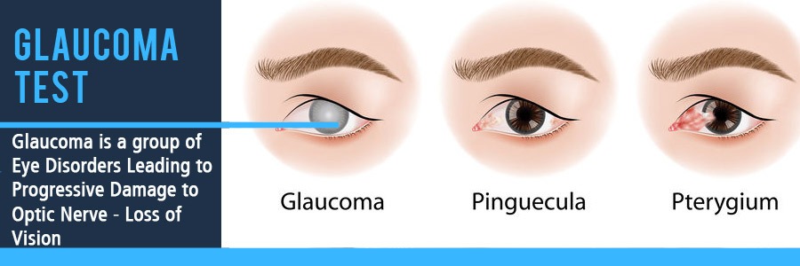
Glaucoma is a group of eye disorders leading to progressive damage to the optic nerve, and is characterized by loss of nerve tissue resulting in loss of vision. Early detection, through regular and complete eye exams, is the key to protecting your vision. Glaucoma can cause blindness if it is left untreated. Unfortunately, approximately 10% of people with glaucoma who receive proper treatment still experience loss of vision.
Glaucoma is the second leading cause of blindness in the world, according to the World Health Organization. Estimates put the total number of suspected cases of glaucoma at over 60 million worldwide.
Blindness from glaucoma is 6 to 8 times more common in African Americans than Caucasians. It is estimated that over 3 million Americans have glaucoma but only half of those know they have it. Call Wohl Optics now to get that Eye and Vision Exam by our optometrist scheduled.
Unfortunately, the vast majority of Americans – 91% - incorrectly believe glaucoma is preventable. Although glaucoma is NOT preventable, if diagnosed and treated EARLY, optometrists can help you control this disease. Medication or surgery can slow or prevent further vision loss. However, vision already lost to glaucoma cannot be restored.To establish a diagnosis of glaucoma, several factors must be present: Because glaucoma is a progressive disease, a change in the appearance of the optic nerve, a loss of nerve tissue, and a corresponding loss of vision confirm the diagnosis. Some optic nerves have a suspicious appearance, resembling nerves with glaucoma, but you may have no other risk factors or signs of glaucoma. If this is you, please closely follow with routine, annual comprehensive eye exams to monitor for any changes.
What is a Glaucoma Test?
What is a glaucoma test like?
- Tonometry measures the pressure within your eye. During tonometry, eye drops are used to numb the eye. Then we use a device called a tonometer to measure the inner pressure of the eye. A small amount of pressure is applied to the eye by a tiny device or by a warm puff of air. The range for normal pressure is 12-22 mm Hg (“mm Hg” refers to millimeters of mercury, a scale used to record eye pressure). Most glaucoma cases are diagnosed with pressure exceeding 20mm Hg. However, some people can have glaucoma at pressures between 12 -22mm Hg. Eye pressure is unique for each person.
- Ophthalmoscopy is a diagnostic procedure that helps your eye doctor examine your optic nerve for glaucoma damage. Eye drops are used to dilate the pupil so that the doctor can see through your eye to examine the shape and color of the optic nerve. The doctor will then use a small device with a light on the end to light and magnify the optic nerve. If your intraocular pressure is not within the normal range or if the optic nerve looks unusual, your doctor may ask you to have one or two more glaucoma exams: perimetry and gonioscopy.
- Perimetry is a visual field test that produces a map of your complete field of vision. This test will help a doctor determine whether your vision has been affected by glaucoma. During this test, you will be asked to look straight ahead and then indicate when a moving light passes your peripheral (or side) vision. This helps draw a "map" of your vision.
- Gonioscopy is a diagnostic exam that helps determine whether the angle where the iris meets the cornea is open and wide or narrow and closed. During the exam, eye drops are used to numb the eye. A hand-held contact lens is gently placed on the eye. This contact lens has a mirror that shows the doctor if the angle between the iris and cornea is closed and blocked (a possible sign of angle-closure or acute glaucoma) or wide and open (a possible sign of open-angle, chronic glaucoma).
Types of Glaucoma
Primary open-angle glaucoma: This is the most common form of glaucoma. One theory is that glaucoma is thought to develop when the eye’s drainage system becomes inefficient over time. This leads to an increased amount of fluid and a gradual buildup of pressure within the eye. Other theories of the cause of the optic nerve damage include poor perfusion, or blood flow, to the optic nerve. Damage to the optic nerve is slow and painless and a large portion of vision can be lost before vision problems are noticed.
Angle-closure glaucoma: This type of glaucoma, also called closed-angle glaucoma or narrow angle glaucoma, is a less common form of the disease. It is a medical emergency that can cause vision loss within a day of its onset. Angle-closure glaucoma can be chronic (progressing gradually) or acute (appearing suddenly). The acute form occurs when the iris completely blocks the drainage of the aqueous fluid. Although an acute attack often affects only one eye, the other eye may be at risk of an attack as well.
Secondary glaucoma: This type of glaucoma occurs as a result of an injury or other eye disease. It may be caused by a variety of medical conditions, medications, physical injuries, and eye abnormalities. Infrequently, eye surgery can be associated with secondary glaucoma.
Normal-tension glaucoma: In this form of glaucoma, eye pressure remains within what is considered to be the “normal” range, but the optic nerve is damaged nevertheless. Why this happens is unknown.
About Wohl Optics Vision Care
Proper eyewear prescription AND fit are vital for your best vision. Fit is something that you will never get right with an online optical business. Veteran owned and operated - best in Bucks County Optical eye care shop.
How could your vision be better? What situations do you feel give you trouble when wearing eyeglasses? That is why we are here.
Optical Services, Products and more
Exclusive Discounts
Military, Veterans, First Responders, Police, Firefighters, Ambulance all receive exclusive discounts (not combined with insurance or other discounts).
We accept most major Vision Insurance Plans.
(215) 443-7706 Phone
(215) 443-8795 FaxWohl Optics
550 Street Rd.
Warminster, PA 18974Veteran Helping Veterans - Local Bucks County, PA Independent Optician

Copyright © 2016-2022
All Rights ReservedCredit Cards Accepted

Private Consultations for your Family: Flexible hours at your convenience by appointment ONLY
Wohl Optics Regular Schedule:
Monday 10:00AM - 5:00PM Tuesday 10:00AM - 4:00PM Wednesday 10:30AM - 6:00PM Thursday 10:30AM - 7:30PM Friday 10:00AM - 6:00PM Saturday 10:00AM - 1:00PM We are offering personal appointments to everyone for the selection of eyeglasses and eyeglass adjustments. We will make every effort to accommodate your schedule. Let us know if you would like to meet at Wohl Optics outside of the above regular scheduled hours.
Service Areas
Bucks County, PA; Montgomery County, PA; Chester County, PA; Philadelphia, PA; Warminster, PA; Ivyland, PA; Warrington, PA;Furlong, PA; Warrington, PA; New Hope, PA; Southampton, PA; Bensalem, PA; Northampton, PA; Hatboro, PA; Willow Grove, PA; Huntingdon Valley, PA; Horsham, PA; Lansdale, PA; Montgomeryville, PA; Newtown, PA; Langhorne, PA; Lahaska, PA; Buckingham, PA; Yardley, PA; Chalfont, PA; Richboro, PA; Doylestown, PA; Glenside, PA; Ambler, PA; Fort Washington, PA; Churchville, PA; Norristown, PA; Washington Crossing, PA; Philadelphia, PA.
Wohl Optics 550 Street Rd. Warminster, PA 18974 (215) 443-7706 Privacy Policy HIPAA Sitemap